Scholl Lab
Understanding how synaptic networks shape visual computation, from single dendrites to behavior.
Understanding how synaptic networks shape visual computation, from single dendrites to behavior.
We study synaptic networks — the vast collection of inputs each neuron receives, and how they drive, suppress, and reshape what a cell computes.
Every neuron integrates a wide collection of inputs that can drive, suppress, or subtly modulate its activity. The operations a neuron can perform are defined by these inputs and their dynamics. Outside of moment-to-moment network dynamics, the primary way a neuron dramatically changes its operational capacity is through plasticity of these inputs — during learning and development. To understand how individual neurons and circuits transform information, we must first build a fundamental understanding of synaptic networks. We believe this knowledge will extend beyond the fundamentals and lead to new insight into circuits underlying neurological and developmental disorders.
Our work uses a variety of techniques to study sensory processing within single cells and across large-scale populations in vivo — historically through electrophysiology (intracellular and extracellular recordings) and multiphoton calcium imaging. We have since expanded into two-photon optogenetics, functional connectomics with electron microscopy, gene editing to disrupt naturally expressed proteins and receptors, and the study of natural behavior.
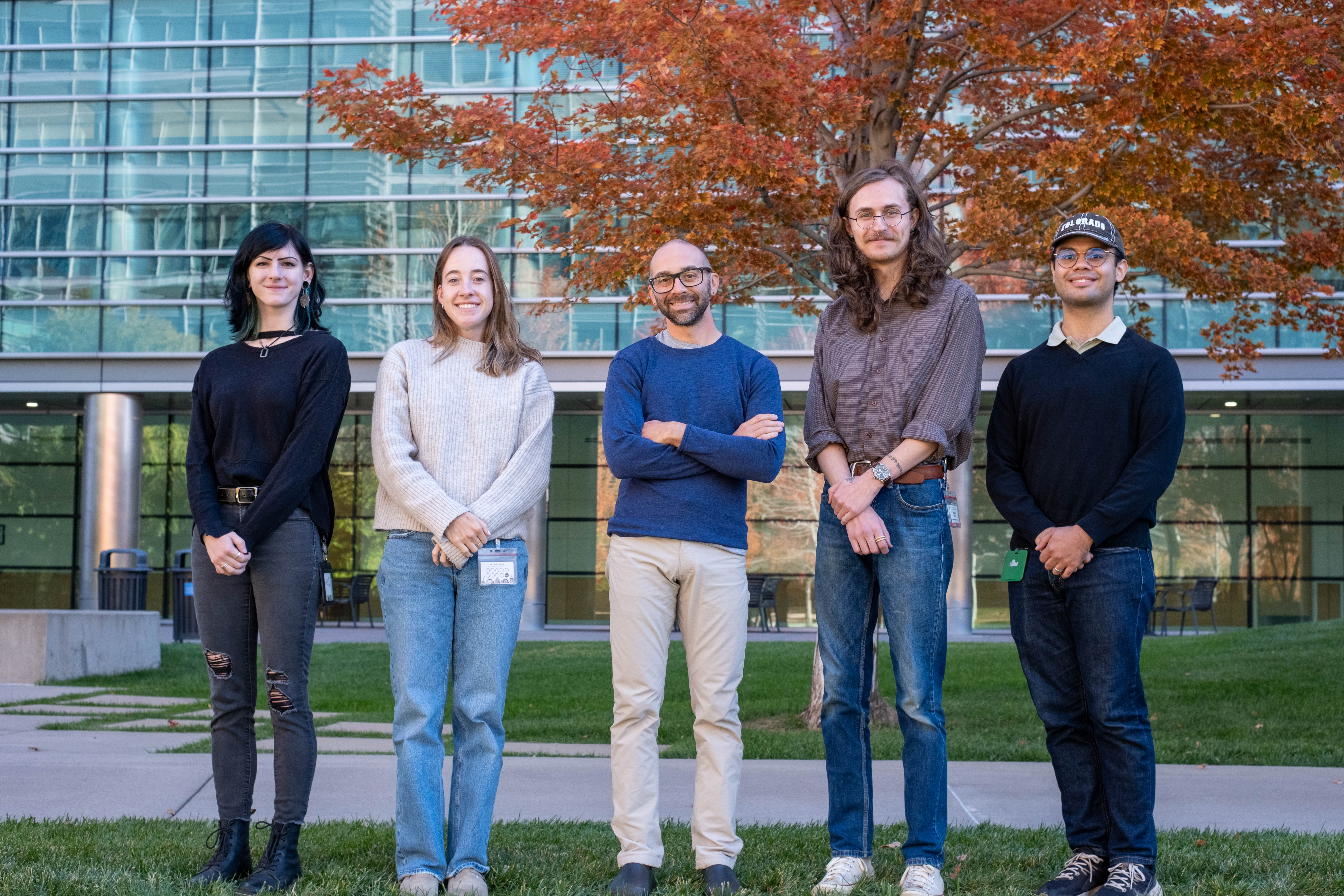
All people deserve to be treated equally, with dignity and respect. Our lab welcomes and encourages individuals from all backgrounds, and we are committed to maintaining a supportive environment. Together, we strive for scientific excellence while learning and honing a variety of skills in the process.
We are part of the Visual Cluster at CU Anschutz, alongside Drs. Gidon Felsen, Alon Poleg-Polsky, and Dan Denman. Our environment provides plenty of resources to share with interested students, trainees, and staff scientists.
Much of our work is made possible by the shared resources and support at CU Anschutz — including the Neurotechnology Center (NTC) and its Optogenetics and Neural Engineering Core, as well as the dedicated animal support staff in the RC1 vivarium. We are grateful for their expertise and care.
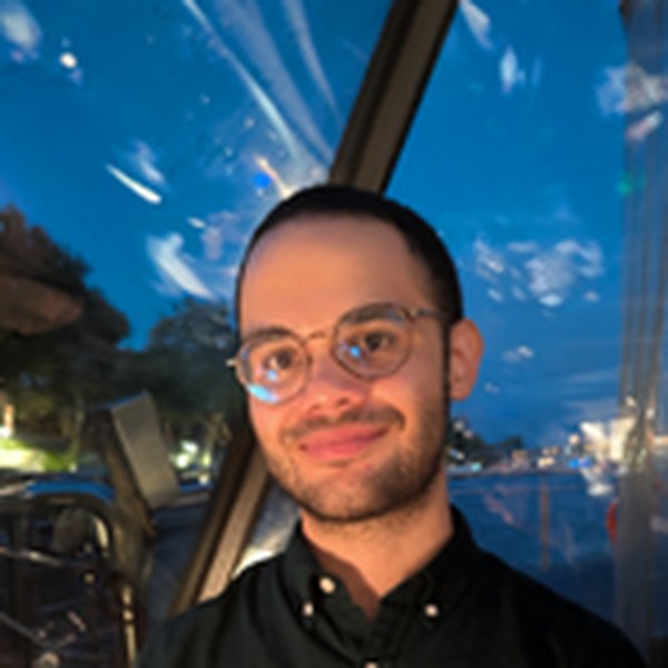
Ben leads the lab's efforts to understand the synaptic basis of visual computation. His work combines in vivo physiology, optical imaging, and connectomics to map how individual neurons integrate their inputs. Before starting the lab at CU Anschutz, he trained at the Max Planck Florida Institute for Neuroscience and the University of Texas at Austin.
How do selective visual responses emerge from a neuron's myriad synaptic inputs? We aim to identify the principles of functional connectivity within layer 2/3 of ferret primary visual cortex — testing the idea that inter-columnar connections following functional-similarity rules are essential for feature selectivity. Using all-optical interrogation, we perturb and map excitatory and inhibitory interactions within and between orientation columns; with two-photon calcium imaging, we map how long-range inputs are organized across the dendritic tree and dynamically recruited to shape somatic responses. This work seeks new computational circuit motifs in ferret V1 — advancing our understanding of cortical circuits and informing brain-inspired AI.
Experience shapes the developing brain — but experience itself changes as the brain matures. Studying ferrets developing naturally, we track how visually guided behaviors (movement, eye position, posture) emerge alongside the maturing circuits that support them. We are now bringing neural recordings into freely behaving animals — combining miniaturized 1-photon (miniscope) and 2-photon (mini2P) imaging, Neuropixels, and markerless 3D pose tracking (FreeMoCap) — to measure active visual processing as it unfolds during natural behavior.
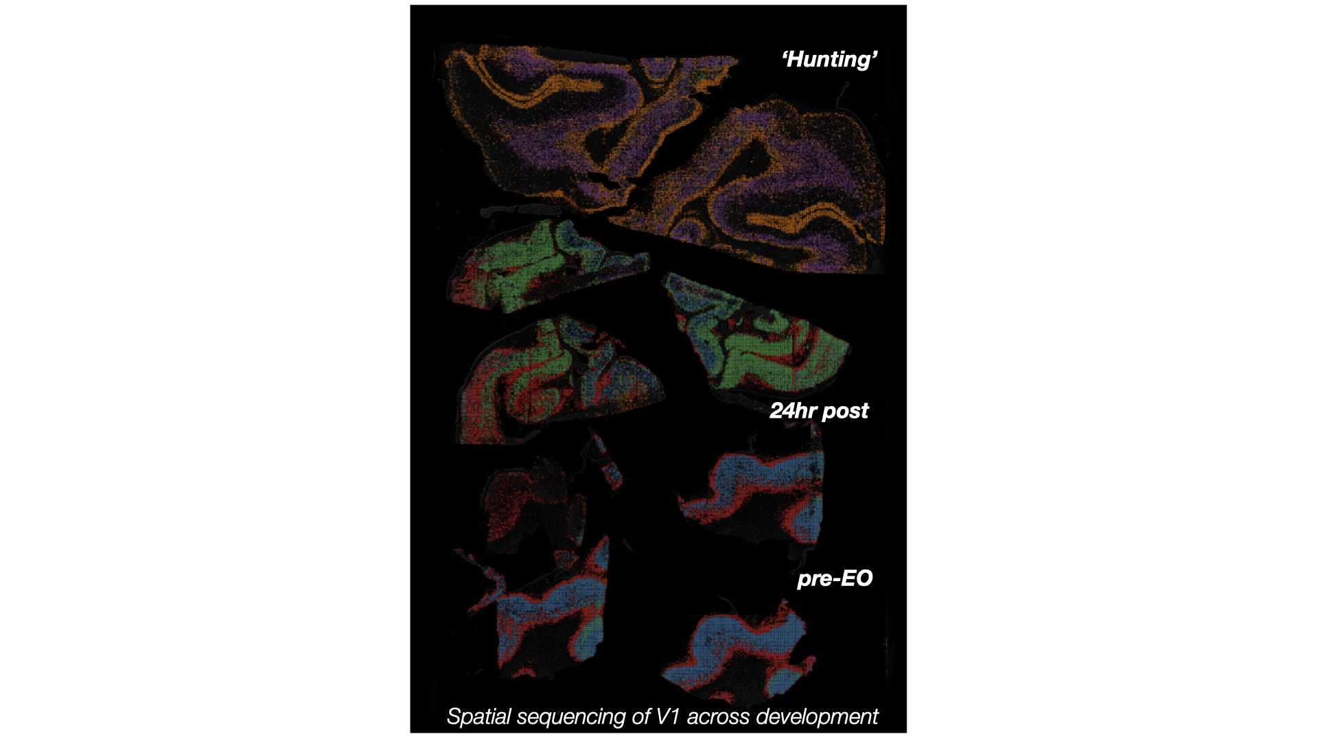
We are building an open molecular resource of the developing ferret brain, combining spatial transcriptomics and single-nucleus RNA sequencing across postnatal development and around eye opening. Preserving spatial context alongside single-cell resolution lets us capture how molecular programs evolve across cortical layers and visual areas as circuits mature. Our goal is a shareable ferret cell-type atlas — a foundation for understanding the emergence of visually guided behavior — with the datasets released to the community as the work is published.
How do motor commands reshape visual processing? Saccades offer an ideal framework — coupling a well-defined motor command to rapid, predictable changes in retinal input — yet the circuits that generate saccadic modulation remain unresolved. The superior colliculus is uniquely positioned to address this, combining sensorimotor circuitry with genetically accessible visual cell types. Using head-fixed mice, high-speed eye tracking, and Neuropixels and two-photon recordings, we dissect how suppression and enhancement during saccades keep perception stable as the eyes move. This work anchors a collaborative Visual Cluster R01.
We collaborate with labs around the world — from image processing and analysis to modeling cortical circuits with real experimental data. Collaborators include Matthias Kaschube (FIAS), Alexander Huk (UCLA), Gordon Smith (UMN), Tal Laviv (Tel Aviv University), Jonathan Matthis (FreeMoCap), Jacob Yates (Berkeley), and Brock Grill (Seattle Children's).
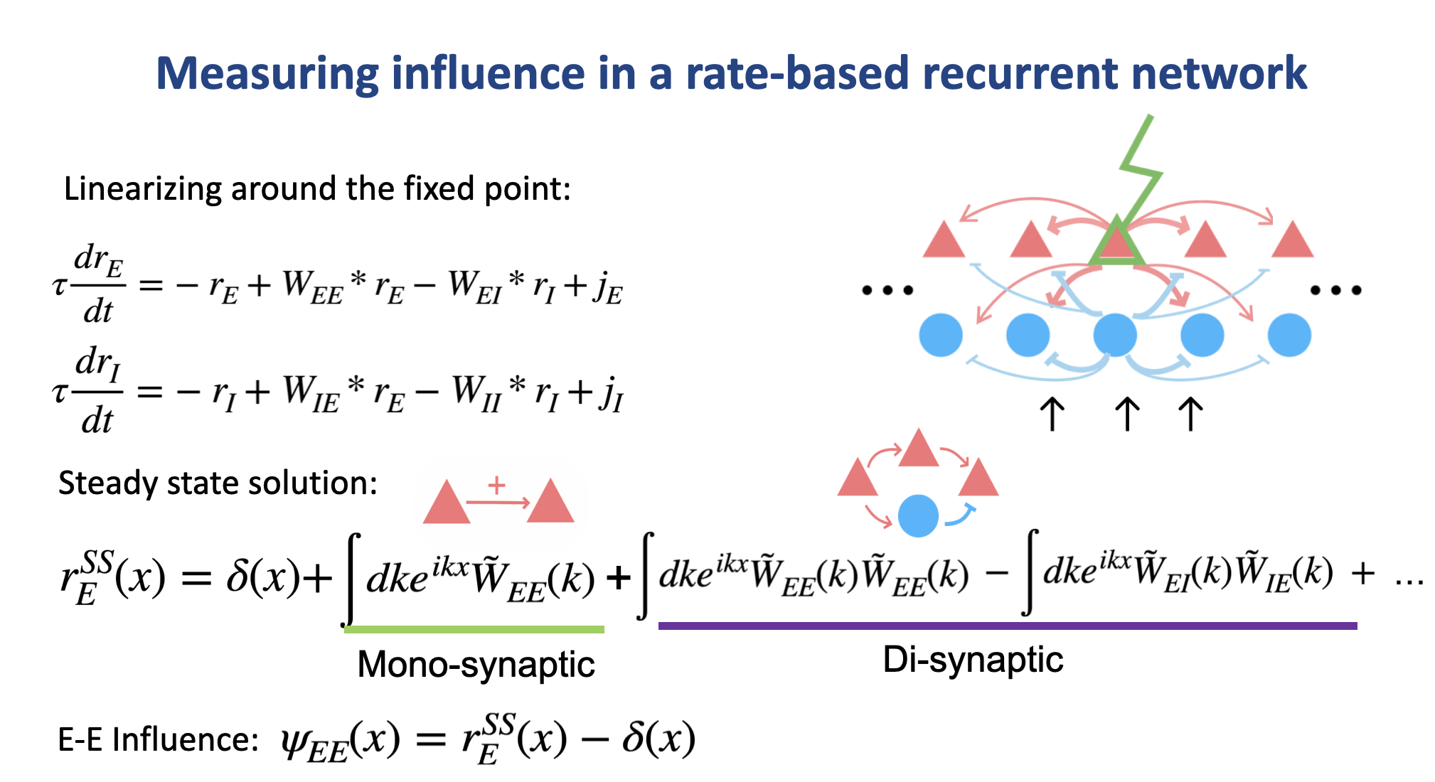
Principles of cortical interactions in modular recurrent networks
In revision · Nature Communications · 2026
View publication →
Fluorescence lifetime-based biosensor for monitoring compartmentalized autophagy dynamics in the intact mammalian brain
In revision · Neuron · 2026
View publication →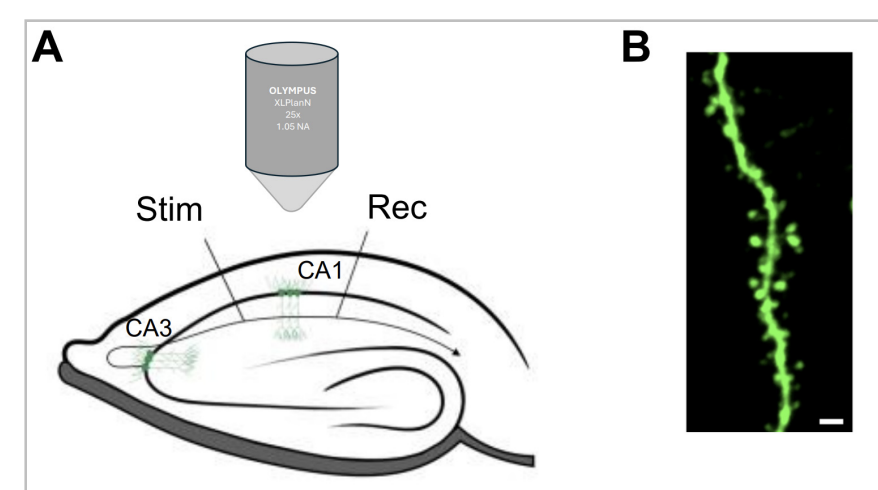
Activation of CP-AMPARs is required for homosynaptic and heterosynaptic structural LTP in the hippocampus
bioRxiv · 2026
View publication →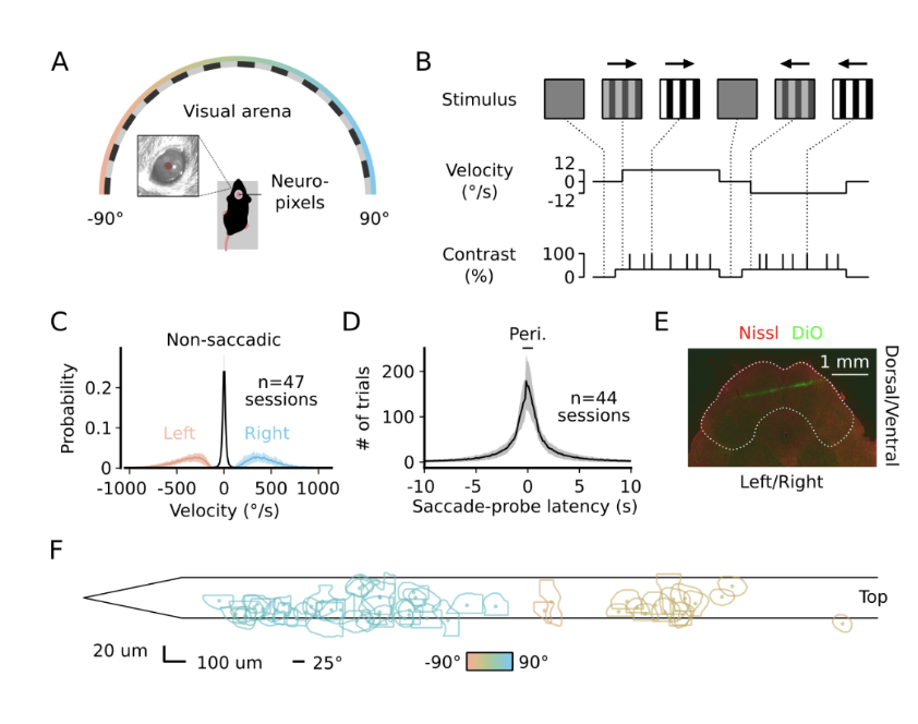
Neural substrates for saccadic modulation of visual representations in mouse superior colliculus
PNAS · 2025
View publication →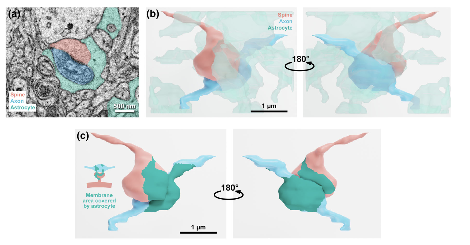
Astrocyte coverage of excitatory synapses correlates to measures of synapse structure and function in ferret primary visual cortex
GLIA · 2024
View publication →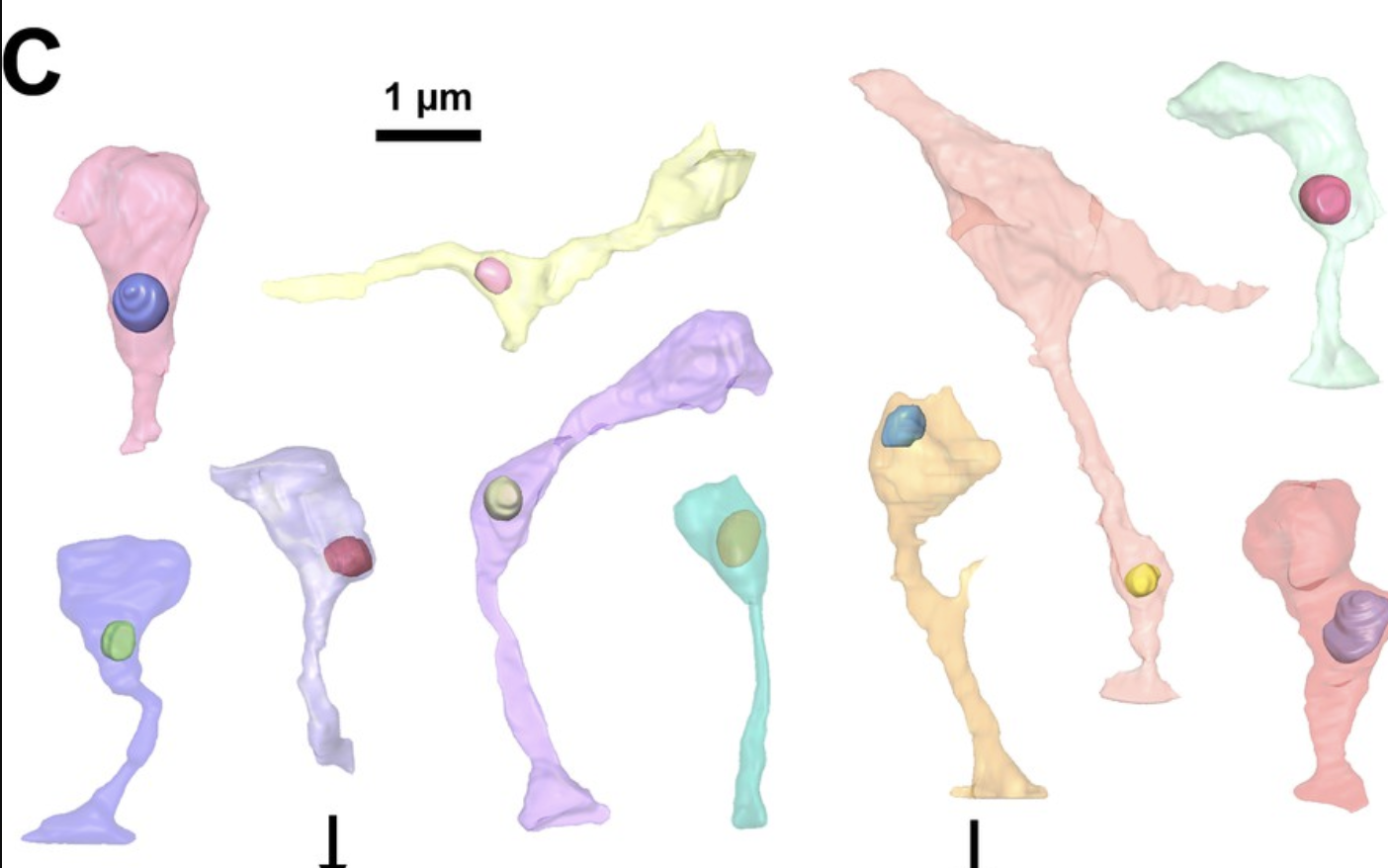
Postsynaptic mitochondria are positioned to support functional diversity of dendritic spines
eLife · 2023
View publication →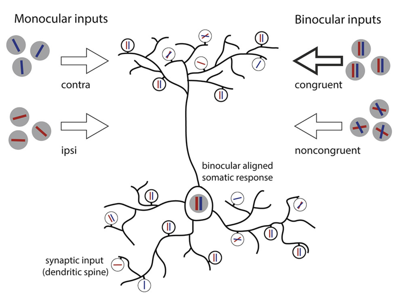
A binocular synaptic network supports interocular response alignment in visual cortical neurons
Neuron · 2022
View publication →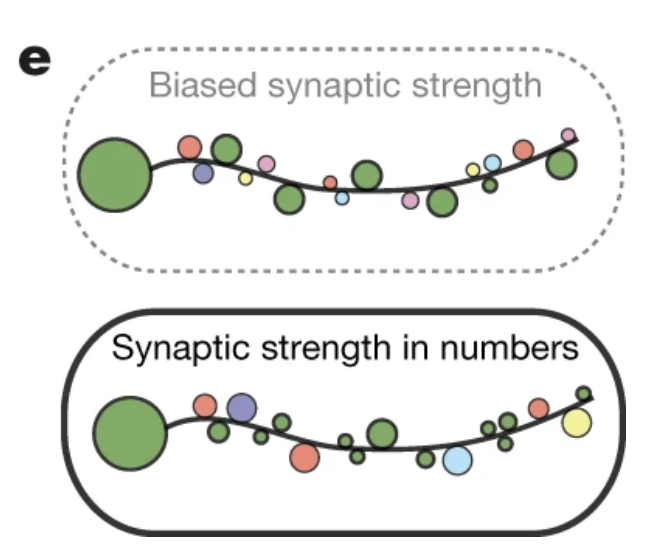
Cortical neuron response selectivity derives from strength in numbers of synapses
Nature · 2021
View publication →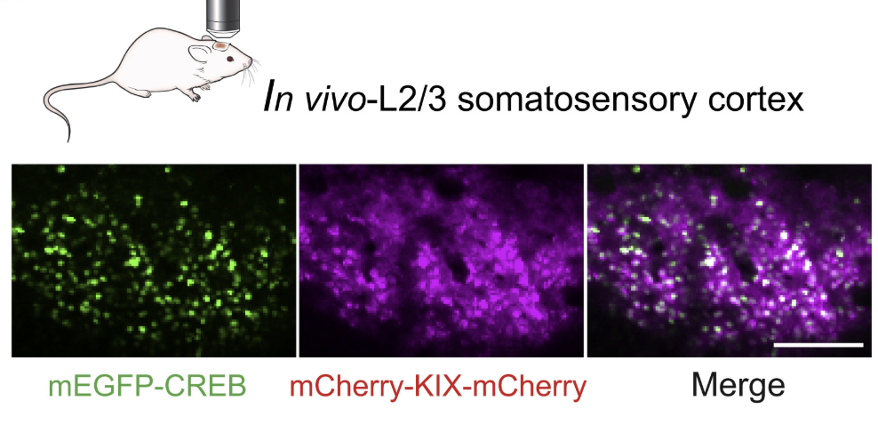
In vivo imaging of the coupling between neuronal and CREB activity in the mouse brain
Neuron · 2019
View publication →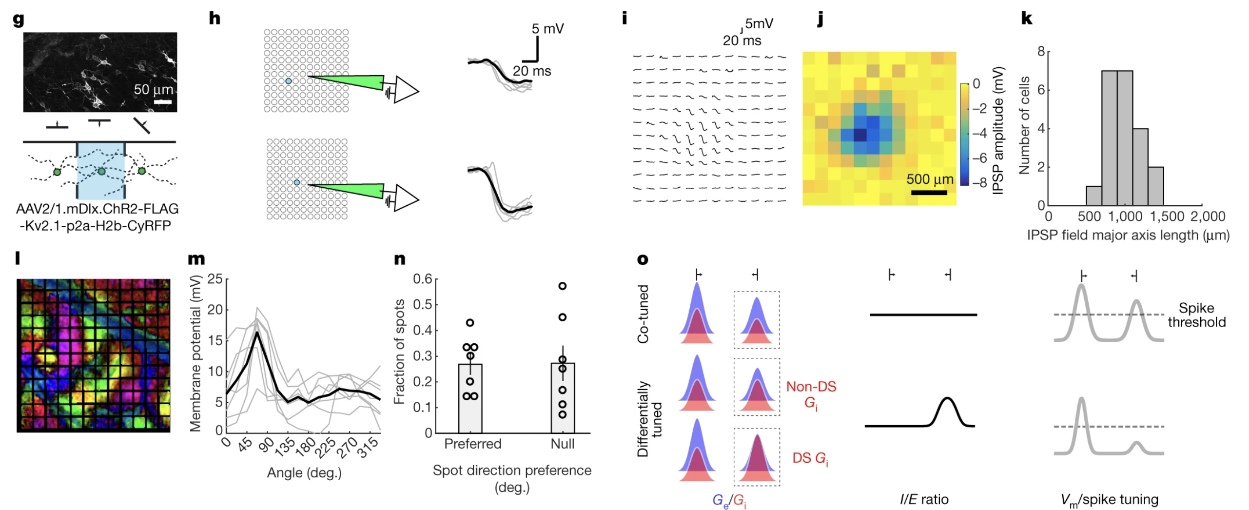
Differential tuning of excitation and inhibition shapes direction selectivity
Nature · 2018
View publication →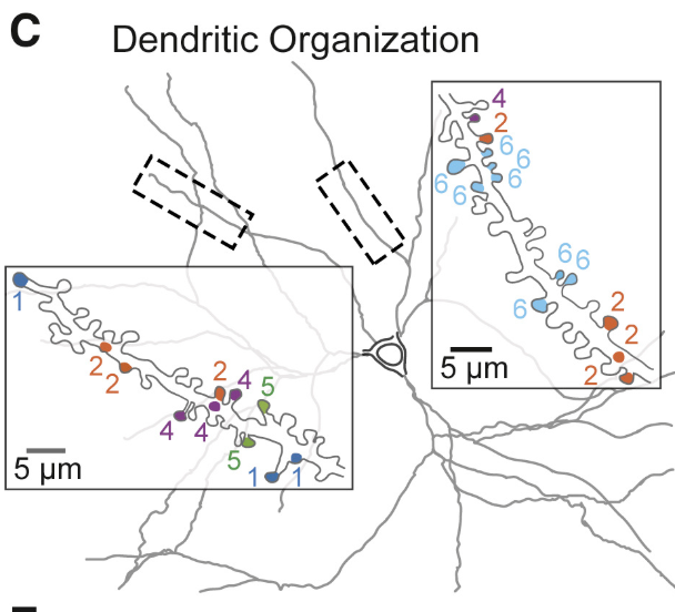
Local order within global disorder: synaptic architecture of visual space
Neuron · 2017
View publication →
Studies the synaptic basis of visual computation using in vivo physiology, imaging, and connectomics.

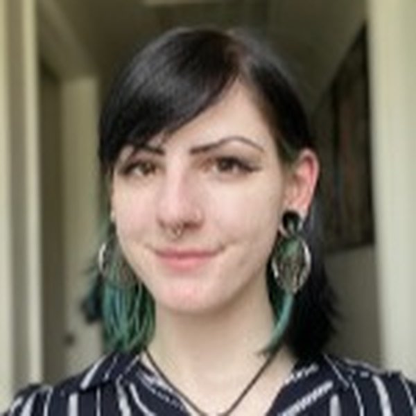
Penny earned her PhD in the Huk lab at UCLA, where she studied how the primate visual system encodes motion and depth to support 3D perception, combining psychophysics with neural recordings. She joins the lab as a postdoc to investigate the synaptic and circuit basis of visual computation.
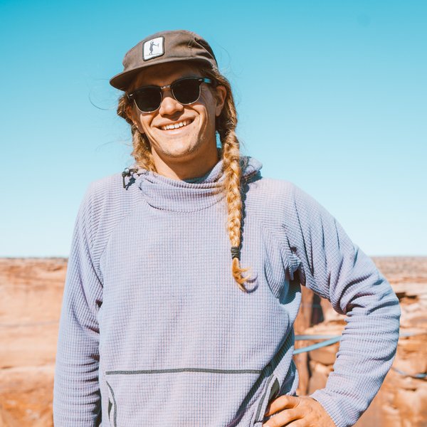
Joe is a PhD student in the Neuroscience Graduate Program (NSP). He has been with the lab since its founding at the University of Pennsylvania and contributes broadly across projects — including co-authoring the Hunt et al. (PNAS) and Kong et al. studies.
Emily is a PhD student in the Neuroscience Graduate Program (NSP).
Philip is a PhD student in the Computational Bioscience Program (CPBS), joining the lab August 1, 2026. He develops software for markerless motion capture (FreeMoCap) and studies natural behavior and neural dynamics.

Rohit is a software engineer interested in building closed-loop behavioral paradigms for studying visual processing and natural behavior.
A research professional previously in the Felsen lab who works closely with our group as part of the Visual Computation Cluster, and a co-author on our collaborative biosensor study with the Laviv lab (Maman et al.).
A research professional previously in the Cruz-Martín lab who supports many projects across our lab and the Visual Computation Cluster; she earned her bachelor's at CU Boulder.
Visual Cluster · CU Anschutz
We are always interested in motivated, curious people who want to understand how synaptic networks shape brain function and behavior. The lab offers a collaborative environment and the chance to learn a wide range of experimental and computational techniques.
Ben trains students through three CU Anschutz programs: the Neuroscience Program (NSP), the Computational Bioscience Program (CPBS), and the Medical Scientist Training Program (MSTP). Feel free to reach out before applying to discuss your background and interests.
We welcome candidates with a strong background in neuroscience, systems physiology, or a related field. Experience with in vivo electrophysiology, optical imaging, or computational analysis is a plus.
We occasionally host undergraduate researchers interested in gaining hands-on laboratory experience.
To inquire, please send a brief email describing your background and interests to benjamin.scholl@cuanschutz.edu.